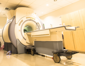
There is increasing evidence that obstructive sleep apnea (OSA) is a heterogeneous disorder with distinct phenotypes that encompass risk factors other than body weight alone. These risk factors include inflammation and trauma to the upper airway, nasal obstruction, craniofacial structure, and the threshold for upper airway collapsibility in response to the negative intraluminal pressure created by apneic events. Dynamic MRI, applied along with recordings of respiratory airflow, upper airway muscle activity via electromyography (EMG), and sleep-wake state via electroencephalography (EEG), can be a potentially powerful tool for examining the anatomical and functional differences in the airways of patients with OSA. A better understanding of the functional mechanisms underlying upper airway dynamics will enable us to develop improved predictive models of respiratory control during natural sleep and during wake-sleep transitions.
(Lead Investigator: Krishna Nayak)
Selected publications:
Seeing sleep: dynamic imaging of upper airway collapse and collapsibility in children.
Nayak KS, Fleck RJ. IEEE Pulse. 2014 Sep-Oct;5(5):40-44. PMID: 25264692
Real-time 3D magnetic resonance imaging of the pharyngeal airway in sleep apnea.
Kim YC, Lebel RM, Wu Z, Ward SL, Khoo MC, Nayak KS. Magn Reson Med. 2014 Apr;71(4):1501-10. PMID: 23788203
Dynamic 3-D MR Visualization and Detection of Upper Airway Obstruction During Sleep Using Region-Growing Segmentation.
Javed A, Kim YC, Khoo MC, Ward SL, Nayak KS. IEEE Trans Biomed Eng. 2016 Feb;63(2):431-7. PMID: 26258929
Evaluation of upper airway collapsibility using real-time MRI.
Wu Z, Chen W, Khoo MC, Davidson Ward SL, Nayak KS. J Magn Reson Imaging. 2016 Jul;44(1):158-67. PMID: 26708099
Real-Time Magnetic Resonance Imaging. Nayak KS, Lim Y, Campbell-Washburn AE, Steeden J. J Magn Reson Imaging. 2022 Jan;55(1):81-99. doi: 10.1002/jmri.27411.
